
Get a Better Understanding of Your Teeth
The anatomy of a tooth is so much more than just white enamel and an awkward gap if you lose one. Let’s dive into this intricate marvel, each layer possessing its unique attributes that work seamlessly together to serve our daily needs like eating and speaking!
What Is a Tooth Made Of?
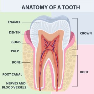
A tooth consists of four main layers: enamel, dentin, cementum, and dental pulp. The enamel is the hard, protective outer layer that covers the crown (visible part) of the tooth. Dentin is the softer layer beneath the enamel that makes up most of the tooth’s structure. The cementum covers the root surface of the tooth and anchors it to the jawbone through connective tissue fibers called periodontal ligaments. Finally, the dental pulp contains nerves and blood vessels that supply nutrients to the tooth. Understanding these components can help promote good oral hygiene habits and aid in the early detection and treatment of dental issues.
Anatomy Overview and Teeth Type Classification
Teeth are an essential part of human anatomy. They serve not only a functional purpose but also have an impact on one’s overall appearance.
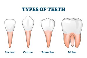
The human mouth contains different types of teeth classified into four groups:
- Incisors: They occupy the front of the mouth and are responsible for biting and cutting food.
- Canines: These lie next to the incisors on both sides of the mouth and are used for biting, tearing, and holding food
- Premolar: They sit between the canines and molars and function primarily as crushing teeth that grind food.
- Molars: Found at the back of the mouth and have a flattened structure ideal for grinding down food.
Each type of tooth performs a specific mechanical action, allowing for efficient food breakdown before swallowing. The position of each tooth can also impact speech production. For example, we rely on our front teeth (incisors) when pronouncing certain sounds such as “f” and “v.”
The number and types of teeth vary from person to person due to medical conditions such as hypodontia or supernumerary teeth; a dentist can examine patients’ dental charting to identify these abnormalities.
Incisors
Incisors contribute significantly to how you project your smile. They give an aesthetic look to your face by determining how much visible gum shows in the top front teeth. The width of the maxilla and mandible, the curvature at the corners of your mouth, and the thickness of the upper lip are all contributing factors in this regard.
Canines
Canines are the most prominent teeth on either side of the upper and lower jaw. Their pointed shape is ideal for biting into food. They also hold a significant role in speech production as they aid in enunciating words such as “she” and “be.”
Premolars
The premolars are present between the canine and molar teeth, with one or two roots depending on their location in the mouth. These teeth assist in chewing food before swallowing it.
Molars
Lastly, molars are located at the back of your jaw. They have a flat surface that enables them to crush food into smaller pieces while enabling you to chew more efficiently. It’s essential to understand that when compared to other types of teeth, molars tend to have more roots to support their larger size.
 The Surface Layer: Enamel and Dentin
Enamel may seem like a shiny, impenetrable layer, but it plays an incredible role in protecting teeth from decay and damage. It’s the hardest substance in the human body. However, enamel isn’t a living tissue; once it’s damaged or lost due to wear and tear or cavities, it can’t regenerate. That’s why taking proper care of our enamel through regular brushing, flossing, and dental check-ups is crucial.
The enamel can have different thicknesses and shapes depending on the type of tooth it covers. For example, incisors have flat, thin enamel on their biting edges since they mainly cut through food. Molars have thicker, more complex enamel cusps and grooves to facilitate grinding and crushing food. Canines have pointed enamel which aids in puncturing food.
Dentin is the layer beneath the enamel that makes up most of the tooth structure. It’s softer than enamel but still forms a protective shield around the tooth pulp. Dentin comprises 70% minerals and 30% organic matter, like collagen fibers. It also contains microscopic tubules that connect directly to the pulp chamber and carry nutrients and fluid to the tooth.
When we eat foods containing sugar and starches, bacteria in our mouth produce acid that attacks our tooth enamel by breaking down its minerals. Over time, this can weaken enamel and cause cavities to form. Brushing with fluoride toothpaste helps remineralize (rebuild) weakened areas of enamel before they become cavities or further damage occurs.
Some people might believe that once tooth enamel is gone or damaged beyond repair, there’s nothing dental professionals can do besides extract the tooth. However, dentists have developed several techniques to replace lost or damaged enamel with artificial materials like tooth-colored fillings, porcelain veneers, or dental crowns.
Tooth Roots
When you think about a tooth, you might only consider the visible crown or surface of it. But there’s much more to explore beneath the gum line. The root of a tooth is an essential component that anchors the tooth into its bony socket within the jaw. The root is composed of two distinct layers — cementum and periodontal ligament.
Cementum is a hard tissue that covers the outer surface of the root. It’s softer than enamel but harder than dentin, and its primary function is to provide attachment for the periodontal ligament fibers that anchor the tooth to the surrounding bone. Cementum also helps in repairing small areas of root damage.
Periodontal ligament (PDL) is a soft connective tissue surrounding each tooth’s root and connecting it to bony sockets within the jawbone. It attaches firmly to the cementum and alveolar bone, allowing for small movements during normal functioning, such as chewing, grinding, and talking.
Features and Function of the Root
The tooth root has several unique features that allow it to function appropriately.
The shape of the root isn’t uniform across different types of teeth — incisors typically have one straight root, while molars have up to three curved roots. The shape is crucial because it determines how well a tooth can anchor itself firmly into its bony socket and how much force it can withstand during normal functioning.
Another feature is root dentin’s presence, which serves as the tooth’s backbone and provides structural support while protecting against breakage. Finally, the blood supply or nerve fibers within the root pulp help detect temperature and pain when triggered by outside stimuli such as cold drinks or food.
Inside the Tooth: Pulp and Root Canal
The pulp is a tissue located in the center of every tooth. It’s comprised of blood vessels, nerves, and cells that help to nourish the tooth. It has a vital role in the formation of dentin during tooth development. The root canal is a space inside the root of the tooth, which houses the pulp. It’s a pathway for blood vessels, nerves, and other tissues to enter and exit from the pulp chamber.
When dental decay reaches the pulp cavity, it results in inflammation and pain due to pressure on nerves in the area. A perforation or crack on the tooth caused by trauma can also result in bacteria reaching the pulp cavity, leading to infection, inflammation, and extensive damage to the tooth structure.
Dentists use root canal therapy as a treatment option for teeth with infected or inflamed pulp. The procedure involves the removal of infected or dead pulpal tissue by cleaning out the root canal space and shaping it before filling it with an inert material that seals it completely. This process helps to save a tooth that would have been otherwise lost.
Protect Your Teeth
We only get one set of permanent teeth in our lives. Protect your teeth from damage and avoid unwanted dental problems by visiting your dentist twice a year for regular checkups and cleanings. Don’t neglect your oral health or smile any longer!


 The Surface Layer: Enamel and Dentin